Cellular Senescence in Human Tissue
Halted cells that stay metabolically active drive aging across multiple tissues.
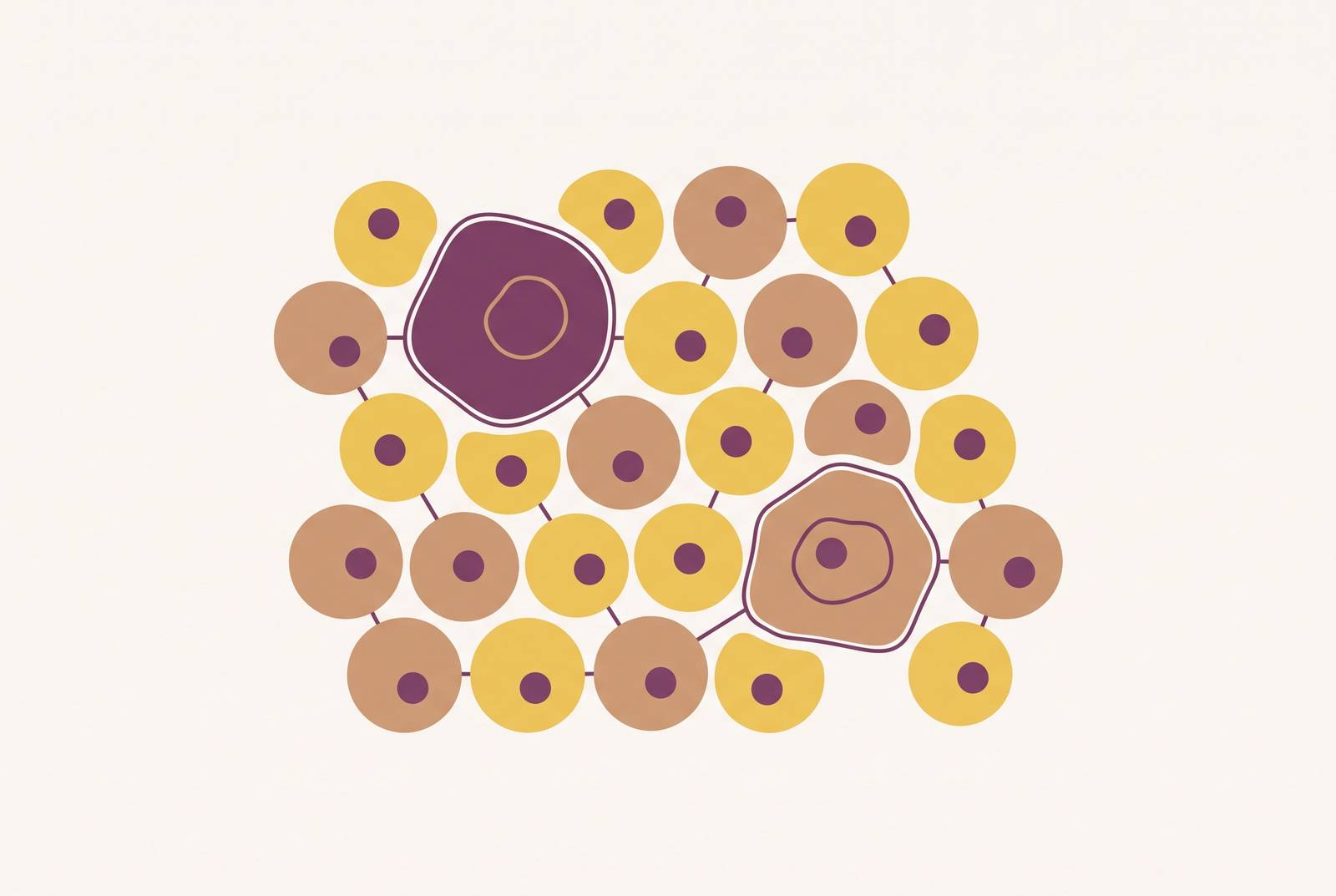
Cellular senescence is what happens when a cell stops dividing for good but doesn't die. It's a permanent halt, not a pause, and the cell sticks around afterward, still metabolically active, still pumping out signals. That single fact, a cell that stops growing but keeps talking, is why aging researchers treat senescence as both a defense mechanism and a slow driver of disease.
The idea traces back to 1961, when two researchers described the phenomenon in human fibroblast cell strains grown in lab dishes. After a set number of divisions, the cells simply stopped, for good. For decades, most of the field treated this as a quirk of tissue culture, something that happened in a dish and probably didn't mean much inside a living body. That assumption held for a long time. Only in more recent decades did senescence get confirmed across multiple species and tissue types in living organisms, not just plastic flasks. Researchers also found that senescence isn't limited to cells that divide: fully mature, postmitotic cells can pick up the same phenotype too.
The current view treats senescence as a shifting, tissue-dependent state rather than a switch that's simply on or off. It counts as one of the formal hallmarks of aging, sitting next to telomere attrition and epigenetic drift, though it gets filed under "antagonistic" hallmarks rather than "primary" ones. That distinction matters. It points to a dual role rather than a straight line from damage to decline. The gap between the 1961 discovery and that acceptance explains a lot about where the science stands now: basic questions about how much senescence exists, in which tissues, and where exactly, are still being worked out.
The two faces of senescence: when halting cell division protects the body
Before senescence became a villain in the aging story, it was doing real work keeping the body safe. Tumor suppression comes first. When a cell picks up an oncogenic mutation, senescence can freeze it in place before it multiplies into something dangerous. That arrest is a built-in brake on cancer, arguably one of the more elegant tricks the body has.
Wound healing is the second role. Senescent cells appear at injury sites on a temporary basis, and their secretions, the same signals that later cause trouble when they overstay, help coordinate repair. Development is the third: senescence programs have been observed during development, playing roles that extend beyond damage response.
What separates helpful senescence from harmful senescence comes down to timing, plain and simple. A healthy immune system clears senescent cells out once they've done their job. Problems start when that clearance slows down or gets overwhelmed, and the cells that were supposed to leave stay put instead.
Calling senescence a double-edged sword isn't just a phrase researchers use to sound clever. The exact same secretory output that recruits immune cells for repair, when it lingers, becomes the thing degrading tissue. Cancer biology makes this plainest: the process that halts a damaged cell from becoming a tumor can, if it drags on, create a chronically inflamed environment that actually favors tumor growth. Same mechanism, opposite outcome, depending entirely on how long it sticks around.
How senescent cells accumulate across a lifetime and why the body cannot clear them fast enough
Cells get pushed into senescence through a handful of well-documented triggers. Telomeres shorten with every division, and once they get too short, that's replicative senescence. DNA gets damaged by radiation, toxins, or oxidative stress. Oncogenes get switched on. And there's stress-induced premature senescence, or SIPS, where oxidative stress is the most common trigger seen in lab studies, often tied to mitochondrial problems: changes in shape, in overall mass, in membrane potential.
A handful of molecular checkpoints run all of this. The p16 pathway arrests the cell cycle in the G1 phase. The p21 pathway can stop things in G1 or G2. The p53 pathway plays a role in SIPS as well. These are the switches that lock a cell into its arrested state.
Why does the pile-up get worse with age? Immune surveillance, the process that normally hunts down and clears senescent cells, weakens as the immune system itself ages. So the very system responsible for taking out the trash slows down right when the trash is piling up fastest.
Then there's the bystander effect, and this is the part that makes accumulation genuinely dangerous rather than just gradual. SASP factors released by one senescent cell can push neighboring healthy cells into senescence too, so the process doesn't just add up, it multiplies. Senescent cells don't quietly fade out either. They stay metabolically active, still secreting, long after they've stopped dividing.
Across studies in multiple model organisms, senescence markers have been found to rise with age, and that general pattern has been observed in multiple tissue contexts. Researchers in aging biology have tied this back to something bigger: the buildup doesn't hit one organ at a time, it stresses multiple tissues at once. That multi-tissue stress is one reason the biology of aging is studied as a systemic process rather than an organ-by-organ one.
What the senescence-associated secretory phenotype does to surrounding tissue
The senescence-associated secretory phenotype, SASP for short, is the cocktail a senescent cell puts out into its surroundings: proinflammatory cytokines, growth factors, matrix metalloproteinases (MMPs), and angiogenic factors.
MMPs do the physical damage. They break down the extracellular matrix, the structural scaffolding that keeps tissue architecture intact. Proinflammatory cytokines feed a slow-burning, chronic state often called "inflammaging," a background hum of immune activity that wears on multiple organ systems over time.
SASP isn't uniform across the body, and that's easy to miss if you picture senescence as one single process wearing different masks. Research using single-cell RNA sequencing has found tissue-specific differences in SASP composition, tied to whether the p21 or p16 pathway dominates in a given tissue. A senescent liver cell and a senescent skin cell aren't necessarily sending the same message.
There's a paracrine effect too. SASP factors reach out and change the behavior of nearby healthy cells, disrupting stem cell niches, slowing tissue regeneration, and dulling normal cell function. It doesn't stop at the local level, either. A 2025 GeroScience study that measured 35 senescence biomarkers in 1,678 participants aged 70 to 79 in the Health ABC cohort found that circulating SASP proteins tracked with higher mortality and worse physical function. That's a systemic signal.
Senescent cells also play defense for themselves. Part of the SASP contributes to immune evasion, suppressing the very immune responses that would otherwise clear them out. A 2024 Nature Aging finding adds another layer: high senescence-related gene activity in the liver correlated with elevated levels of specific proteins detectable in blood, which suggests these cells send signals well past their home tissue.
The tissues that accumulate senescent cells most consequentially, and their clinical presentation
Senescent cells get implicated in nearly every major chronic disease tied to age, which makes this less a tissue-specific story and more a body-wide one. Still, a handful of tissues show the pattern with particular clarity, and skeletal muscle deserves more attention than it usually gets. Senescence in the cells responsible for muscle repair impairs regeneration, and that's a big piece of what drives age-related muscle loss, not just disuse or low protein intake, whatever the supplement aisle wants you to believe.
In adipose tissue, senescent fat cells build up alongside obesity and age, and SASP output from fat tissue drives inflammation that reaches well past the fat itself. In blood vessels, senescent vascular cells contribute to arterial stiffening and atherosclerosis. In the lungs, senescent cells get tied to fibrosis, a loss of elasticity and repair capacity that makes breathing harder over time. In the brain, senescent glial cells have been associated with neuroinflammation and neurodegeneration. In skin, senescent cells in the skin are associated with thinning, collagen loss, and slower wound healing.
Cancer treatment adds a wrinkle. Chemotherapy induces what's called therapy-induced senescence, or TIS, and it can leave patients carrying a senescent cell burden well beyond what's expected for their age. A 2026 Nature Aging study led by Cole and colleagues found that the secretome of therapy-induced senescent ovarian cancer cells drove metastatic spread by reprogramming the metabolism of nearby cancer cells through fructose signaling. A protective treatment mechanism, backfiring in a specific and traceable way.
Frailty shows the connection even before old age sets in. In a study of 543 adults aged 26 to 78 in the Midlife in the United States Refresher cohort, the senescence marker p16INK4a was elevated in whole blood from middle-aged adults already showing signs of frailty and trouble with daily activities. The link between senescence and functional decline doesn't wait for someone to turn 70.
How much is actually known about where these cells live in the body? Less than most people would guess. The NIH's SenNet Program, launched in 2021, exists specifically to map where senescent cells appear, and in what quantity, across human organs and across the lifespan. That a mapping effort like this is still underway says something about how incomplete the current picture really is.
Why measuring senescent cells in the body is harder than it should be
No single marker reliably identifies a senescent cell on its own. That's the core problem, and it explains why so much of the science still moves cautiously. Senescent cells are also rare and scattered through tissue, which makes them genuinely hard to find, let alone count with any precision.
The most widely used marker is SA-β-Galactosidase, or SA-β-gal, which picks up on increased lysosomal activity at pH 6.0 (compared to pH 4.0 in normal cells). It throws false positives, though. Macrophages, dense clusters of cells, and exposure to hydrogen peroxide can all trigger the same signal without any senescence involved.
p16INK4a and p21CIP1 are the two other commonly used markers, both tied to cell-cycle arrest. But single-cell RNA research has found they often don't show up together in the same cells across tissues, and they follow separate timelines. A cell testing positive for one won't necessarily test positive for the other. p21 in particular isn't consistently elevated across senescent cells, which makes it a shaky marker to lean on by itself.
Most casual readers of this field get it backwards here. They assume one good test will eventually settle the difficulty of measuring senescence, the way a blood glucose reading settles a question about diabetes. It won't, because the underlying biology resists it. SenNet's published recommendations call for a tiered approach instead: at minimum, one marker of growth arrest, one SASP marker, and a functional test confirming the cell cycle exit is stable. No single test, however well established, counts as proof on its own.
The deeper issue is heterogeneity. Senescence isn't one uniform program running the same way in every cell. Different tissues host molecularly distinct subpopulations of senescent cells, and newer single-cell and spatial omics tools are only starting to map how varied that landscape actually is. SenNet's stated goal is to eventually build a full blueprint of senescent cells across human organs. That blueprint doesn't exist yet.
What blood-based biomarkers can currently tell us about senescent cell burden
Whole blood RNA sequencing can pick up p16INK4a (CDKN2A) expression, along with DNA damage response markers and SASP factors, and older age consistently tracks with higher levels of all three. p16INK4a checks the boxes researchers look for in an aging biomarker: it rises with age, it tracks with functional decline, and it's detectable in a sample that's easy to collect.
One specific assay, looking at T-cell p16 variant 5, has research support as a way to screen and select participants for senolytic drug trials. Correlated plasma SASP markers offer a less technically demanding stand-in for that same purpose. On the plasma side, the 2025 GeroScience study of 1,678 participants aged 70 to 79 measured 35 senescence biomarkers in baseline blood samples and tied specific levels to mortality risk and physical function outcomes down the line. The 2024 Nature Aging research mentioned earlier found circulating molecules in blood that correlated with liver senescence gene activity, hinting that senescence happening deep in an organ like the liver might be readable from a blood draw.
None of this amounts to a direct headcount of senescent cells sitting in someone's tissue, though. Plasma markers are proxies, useful ones, but proxies all the same. The field is working toward validated panels, and nothing here counts as standard clinical practice yet, no matter what a wellness clinic's marketing brochure implies.
One intervention does have direct evidence behind it: physical activity. Research published in a peer-reviewed gerontology journal found that exercise lowered senescence biomarkers and was associated with a delay in mobility disability. That's a rare case in this field where a specific, testable habit shows a measurable effect on the biology itself, not just on how someone feels day to day.
What does this mean for someone getting routine bloodwork today? Elevated inflammatory markers like IL-6 or CRP may partly reflect SASP activity. Understanding senescence doesn't hand anyone a new test to order, but it adds a layer of meaning to markers already sitting on a standard lab report.
What lifestyle, environmental, and medical exposures accelerate senescent cell accumulation
Oxidative stress appears repeatedly in experimental data as the most common trigger behind stress-induced premature senescence. It connects to diet, air pollution, smoking, and a sedentary lifestyle, the usual suspects wearing a slightly different hat here.
Mitochondrial dysfunction drives it too. Changes in mitochondrial shape, total mass, and membrane potential play a central role in pushing cells toward senescence. Mitochondrial health and senescent cell burden are, in a real sense, the same conversation looked at from two angles.
DNA damage from ionizing radiation, UV exposure, or genotoxic chemicals speeds up the damage-triggered path into senescence. Chemotherapy stands out as one of the strongest accelerants outside of aging itself, and patients who go through it can end up with a senescent cell burden associated with faster biological aging than their chronological age would suggest.
Chronic disease works both directions here, and this is where the loop gets underrated as a target. It speeds up senescent cell accumulation, and the buildup of those cells makes the chronic disease worse in turn. Neither side of that loop is the root cause on its own. Treating either side alone will always leave something on the table.
Epigenetic changes, shifts in histone proteins and broader chromatin remodeling, act as both a trigger for senescence and a marker of it. This is part of how genetics, environment, and daily habits all funnel into the same pathway, and how much senescence burden is visible at a given age reflects a lifetime of accumulated exposure. That means at least part of it sits within someone's control.
The current state of senolytic and senomorphic research for healthy aging
Two main strategies are being tested against senescent cell buildup, and they take opposite approaches to the same problem. Senolytics are drugs built to selectively kill off senescent cells, cutting off SASP at the source. Senomorphics take the gentler route: they suppress the SASP signal itself without killing the cell, muting the inflammatory output while leaving the cell in place.
A third approach looks less like elimination and more like reversal. Partial reprogramming using Yamanaka factors (OCT4, SOX2, KLF4, and MYC, often shortened to OSKM) has, in experimental models, erased senescence markers and restored function in aging tissue when applied briefly, without triggering tumors. That's a genuinely different concept: rejuvenating the cell instead of removing it.
Cancer treatment sits at an interesting crossroads here. Therapy-induced senescence can suppress tumor growth in the short term, but if it lingers, the resulting SASP may push surviving cancer cells toward stem-cell-like traits that raise the risk of relapse. Researchers are exploring senolytics timed to follow chemotherapy specifically, to clean up that lingering senescent burden before it causes new problems.
None of this has reached clinical practice yet, and anyone claiming otherwise is getting ahead of the evidence. Senolytic trials in humans are underway, but no senolytic drug currently has approval for any aging-related use. This is translational science: promising, but not something a doctor prescribes today. Commercial interest is tracking right alongside the science regardless. The longevity and anti-senescence therapy market reached $29.9 billion in 2025, growing at a 6.3% compound annual rate, and that gap between market size and clinical approval says something about how far ahead commercial interest has run.
So what can someone actually do with all of this right now? Physical activity has direct evidence behind it for lowering senescence biomarkers, more than any supplement or device sold under a longevity label today, and that alone should settle which of the two gets the benefit of the doubt. Cutting down on everyday oxidative and genotoxic exposures is backed by the same underlying biology. This includes smoking, excess sun, and pollution where it's avoidable. Tracking inflammatory markers over time offers a rough window into SASP-related burden, even with senolytic therapies still years from the clinic. Senescence research is still filling in basic facts about where these cells live and how many there are. The broad shape of the story is solid enough to act on already. It's protective at first, corrosive once it piles up.
Sources
- SenNet recommendations for detecting senescent cells in different tissues - PMC
- Frontiers | The interplay of cellular senescence and reprogramming shapes the biological landscape of aging and cancer revealing novel therapeutic avenues
- Charting human cellular senescence in aging and disease: Cell
- Biomarkers of Cellular Senescence and Aging: Current State‐of‐the‐Art, Challenges and Future Perspectives - Muthamil - 2024 - Advanced Biology - Wiley Online Library
- Cellular Senescence and Aging: Mechanisms, Disease Convergence, and Therapeutic Frontiers - PMC
- Cellular senescence as a therapeutic target for aging intervention
- Frontiers | Cellular senescence: from homeostasis to pathological implications and therapeutic strategies
- pure.johnshopkins.edu