Mitochondrial Dysfunction as an Aging Driver
Mitochondrial damage triggers a self-amplifying cycle that speeds aging across the body.
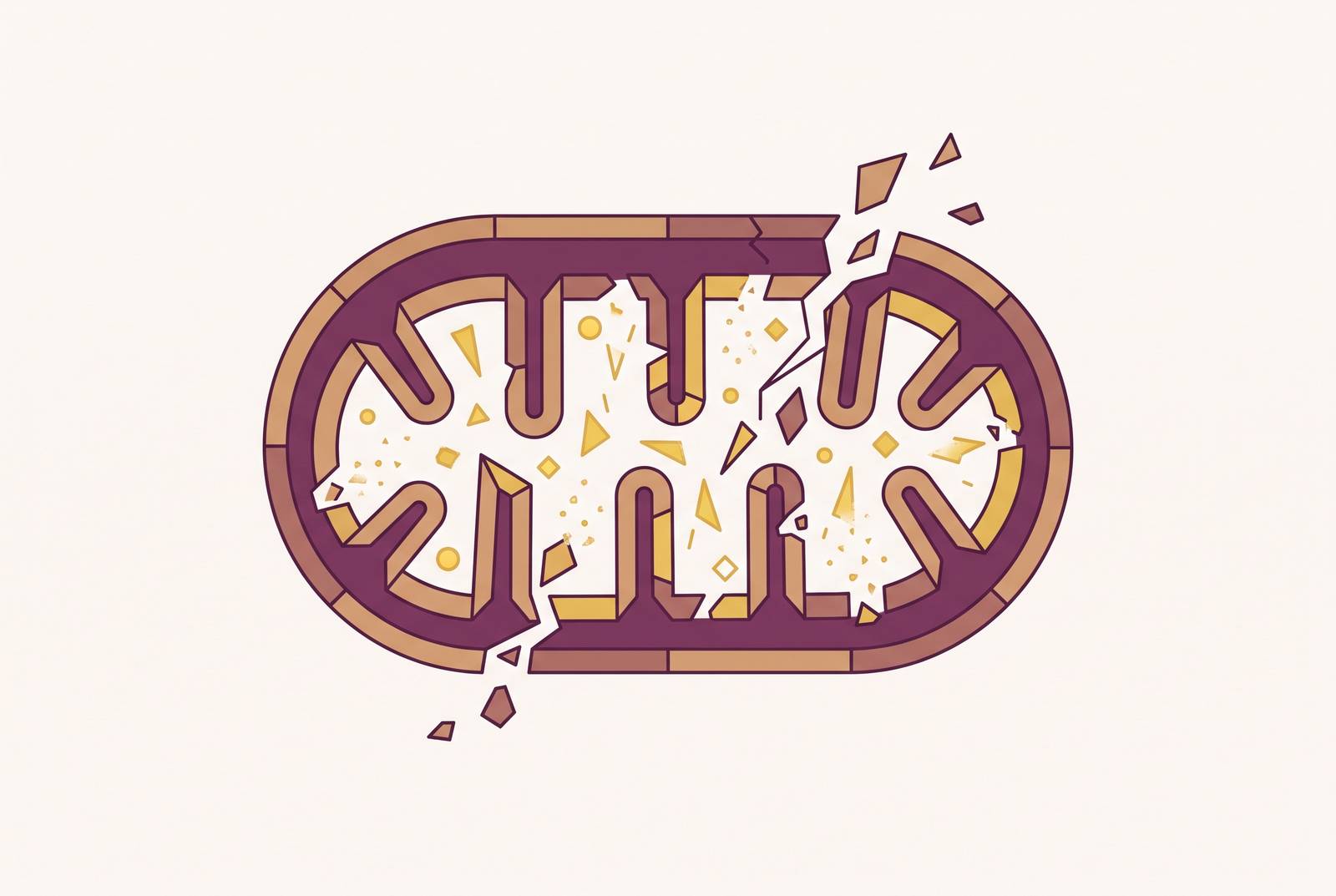
Mitochondrial dysfunction driving aging is visible through five mechanisms. It drives it, through five mechanisms, ROS buildup, mtDNA damage, broken dynamics, failed cleanup, and inflammation, that feed each other and speed decline across the whole body. The 2023 hallmarks of aging paper by López-Otín and colleagues, published in Cell, updated the field's framework to twelve hallmarks. Mitochondrial dysfunction sits among them, and it's not just one item on a list. It's the item that touches the other eleven.
The paper classifies it as an "antagonistic hallmark." That's a fancy way of saying the same response that protects a young cell turns around and wrecks an old one. Whether it helps or hurts comes down to timing and degree, not the response itself. Most people know the line from tenth grade biology: mitochondria are the powerhouse of the cell. True enough, as far as it goes, but thinking, healing a cut, pumping blood, every one of those runs on energy those organelles produce, and that centrality gets underweighted given how much rides on it. What follows traces five mechanisms that lock together, then turns to the organs where the damage becomes apparent.
ROS Accumulation: From Normal Byproduct to Self-Amplifying Damage Signal
Normal energy production works like an assembly line. Glucose breaks down to pyruvate, pyruvate feeds the TCA cycle, that cycle produces NADH and FADH2, and those molecules hand off electrons through the electron transport chain to make ATP. Along the way, some electrons leak. Running the machine generates reactive oxygen species, mainly superoxide anion and hydrogen peroxide, as a side effect nobody can avoid.
Under normal conditions, enzymes like catalase and superoxide dismutase mop this up before it causes trouble. Aging throws that balance off. The mitochondrial free radical theory of aging, first proposed back in 1972, laid out the mechanism: mitochondria produce ROS, that ROS damages mitochondrial components, and the damaged components produce even more ROS. A loop, feeding itself.
The initial ROS was never the real problem. Cells make ROS all the time and handle it fine. What fails is the brake, the antioxidant defense that's supposed to catch the byproduct before it snowballs. Once that brake weakens, the damage stops limiting itself and starts sustaining itself instead.
And ROS doesn't stop at one target. It hits DNA, RNA, proteins, and lipids, laying down molecular wreckage that every other mechanism in this piece builds on. At low levels, ROS actually works as a signaling molecule, helping regulate normal cell processes, so the danger sits in the excess, not the molecule itself. Anyone wondering whether an antioxidant supplement helps or just jams a signal the cell still needs is running straight into that distinction. A 2025 review in Signal Transduction and Targeted Therapy makes a related point: oxidative stress and inflammation, both rooted in mitochondrial dysfunction, cause aging. They don't just trail behind it as symptoms, and that's a meaningful difference if you're trying to figure out where to intervene.
How mtDNA damage accelerates the feedback loop ROS begins
Mitochondria carry their own genome, separate from the DNA sitting in the nucleus. It runs small, just 16.5 kilobases, coding for 37 genes. Damage that genome, and the machine itself takes the hit directly.
Why does mtDNA take more punishment than nuclear DNA? A few reasons stack up. It sits right against the inner mitochondrial membrane, exactly where ROS gets made. It carries no histone coating, the protective wrapping nuclear DNA gets. Its repair systems run weaker than the ones guarding the nucleus.
mtDNA also behaves differently than most people expect. Cells carry many copies of it, a state called polyploidy, and it passes down only through the maternal line. Mutant copies often sit right alongside healthy ones inside the same cell, a state called heteroplasmy. Dysfunction becomes visible only once the mutant fraction crosses a threshold; below that line, a cell can carry real damage and keep humming along like nothing's wrong. That threshold, and how fast the mutant load climbs toward it, varies by tissue, which is a big part of why the brain, heart, and skeletal muscle tend to show age-related decline earlier than other organs.
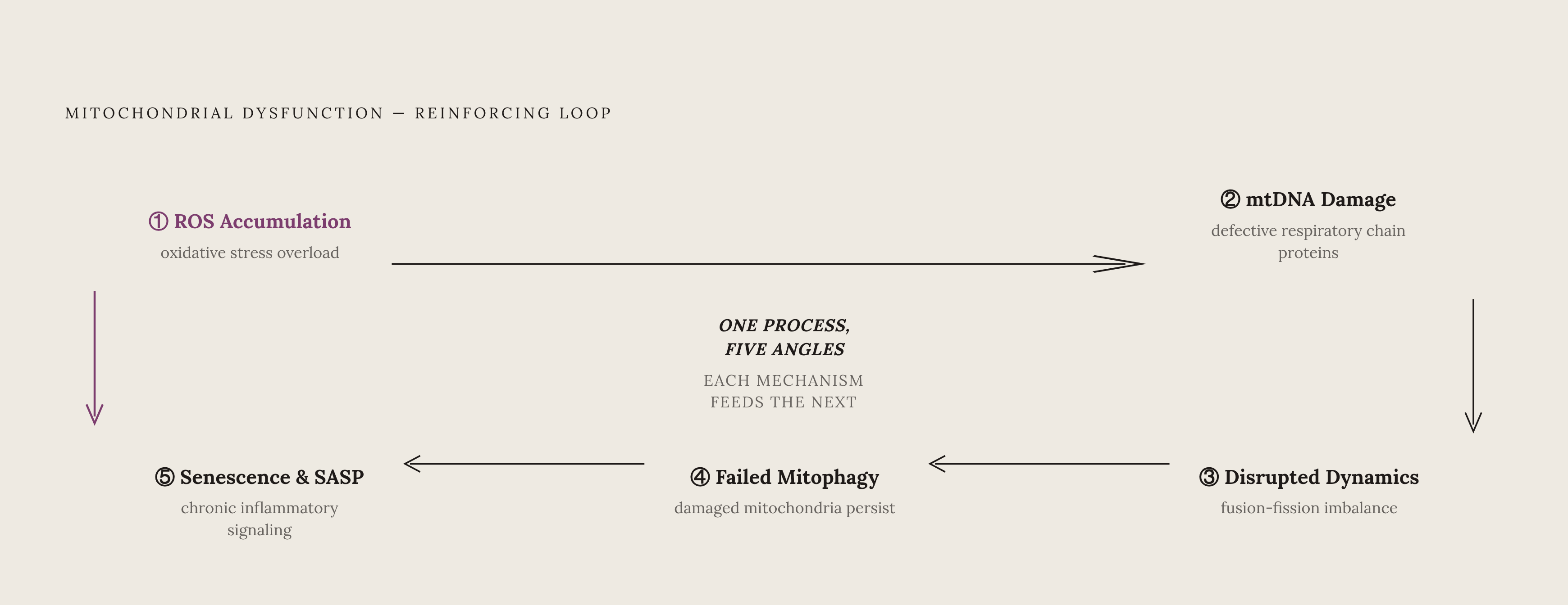
This is where the feedback loop from the ROS section closes. ROS damages mtDNA, damaged mtDNA codes for defective respiratory chain proteins, those defective complexes leak even more electrons, and the leaking produces more ROS. Round and round.
A second route into this same loop runs through telomeres. Telomere DNA damage activates p53 signaling, which suppresses two mitochondrial regulators, PGC-1α and TFAM, both needed to keep OXPHOS and ATP synthesis running. Research has found a connection between nuclear DNA damage and mitochondrial DNA copy number in oxidatively stressed white blood cells, tying two separate hallmarks of aging together at the molecular level.
Heteroplasmy and the threshold effect explain something practical, too. mtDNA damage can build for years without a single outward sign, until the mutant load finally tips over the line and function drops all at once. Catching early biomarkers matters precisely because the symptom becomes visible so late, long after the damage that caused it.
The Effect of Disrupted Mitochondrial Dynamics on Network Health
Mitochondria aren't fixed, solitary blobs sitting in the cytoplasm. They fuse together, merging their networks to share resources and dilute out damage, and they split apart through fission, which segregates damaged units so the cell can flag them for removal. Aging throws this fusion-fission balance off in two directions at once, and both directions cost the cell.
Too much fission leaves behind fragmented mitochondria that run less efficiently. Too little fusion means damage can't get diluted across the network anymore, so defects concentrate instead of spreading thin. Either way, the cell loses its grip on a healthy, well-mixed mitochondrial pool. If you had to pick which failure mode matters more, fission running unchecked is the one to watch: it's the direction most directly linked to the fragmented, inefficient mitochondria that cardiac and neural tissue, the organs that can least afford it, are least equipped to tolerate.
The fallout reaches past ATP output. Disrupted dynamics also throw off calcium handling and mess with amino acid and lipid metabolism. Tissues that run the hungriest for energy feel this fastest: cardiac tissue sees its energetics decline, diastolic dysfunction sets in, and heart failure can follow. Skeletal muscle loses its role as a hub for regulating energy balance and redox chemistry across the whole body. Neurons lose the ability to shuttle mitochondria along axons, exactly where they're needed most.
A 2025 review lays out the mechanism for the heart specifically: oxidative stress damages mtDNA, RNA, lipids, and proteins, and hits cardiomyocyte contractile function directly through the molecular wreckage it leaves behind.
Sex differences are visible here too, and the pattern cuts against a common assumption that aging hits men and women on the same clock. Women often show signs of mitochondrial impairment earlier than men do. Estrogen appears to help regulate mitochondrial gene expression, so its decline during menopause may strip away a layer of protection, raising women's vulnerability to this kind of damage as they age.
Failed Mitophagy and the Accumulation of Damaged Mitochondria
Mitophagy is the cleanup crew: the process that tags worn-out or damaged mitochondria and hands them to lysosomes for breakdown. Aging weakens this system through two failures happening at once, and neither one alone would do nearly the damage that the two together manage.
First, the cell makes fewer new mitochondria to begin with, a downstream effect of the same PGC-1α and TFAM suppression covered earlier. Second, the clearance system itself gets sloppy, letting damaged organelles pile up instead of getting swept out. Research in C. elegans, cited in a special issue of Aging and Disease (Hu et al.), found that a protein called presenilin controls the physical contact points between mitochondria and lysosomes, contact the clearance process depends on. Disrupt that contact, and clearance stalls cold.
What happens when the damaged units stick around instead of getting cleared? They keep leaking ROS and shedding fragments of mtDNA into the cell. Those fragments spill into the cytosol and sometimes the extracellular space, where the immune system reads them as DAMPs, damage-associated molecular patterns, essentially alarm bells with no fire to point to. DAMPs switch on inflammasomes and other immune signaling pathways, and a local cleanup failure turns into a body-wide inflammatory trigger.
Mitochondrial-derived vesicles, small packages that carry mtDNA fragments, metabolites, and proteins out of the cell, activate immune responses wherever they land. This pathway has been linked to Alzheimer's disease and Parkinson's disease.
Skeletal muscle is one tissue where age-related mitophagy failure draws particular attention, and for good reason: it isn't just a local energy problem. When muscle mitophagy machinery breaks down, the fallout ripples into metabolism system-wide. The twelve hallmarks framework lists "disabled macroautophagy" as its own separate hallmark, distinct from mitochondrial dysfunction, but mechanistically the two tangle together closely enough that treating them as separate stories misses half the picture.
Mitochondrial Dysfunction, Cellular Senescence, and Chronic Inflammation
Cellular senescence describes a cell that has permanently stopped dividing but refuses to die. Instead, it sticks around and pumps out a mix of inflammatory signals known as the senescence-associated secretory phenotype, or SASP. These cells build up with age, and mitochondrial dysfunction ranks among the main things that pushes a cell into that state and keeps it stuck there.
A few routes lead there. Weakened OXPHOS efficiency and shifting metabolite levels change a cell's energy and redox balance in ways that favor permanent arrest over normal cycling. Separately, mtDNA that leaks into the cytosol from apoptotic stress or failed mitophagy gets picked up by innate immune sensors, which can drive SASP production directly. Shifting metabolite levels and redox imbalance add further layers, throwing off signals a cell normally relies on to keep cycling on schedule.
Once SASP starts, it doesn't stay contained to the cell that started it. Senescent cells secrete factors that affect neighboring cells, propagating inflammatory signals cell to cell, across a tissue.
That spread is also where chronic inflammation enters the story. Mitochondrial dysfunction triggers inflammation through DAMPs, inflammasome activation, and recruitment of inflammatory cells, and oxidative stress and inflammation feed each other in both directions, a cycle that keeps damaging the tissues around them. This low-grade, persistent state, sometimes called "inflammaging," sits upstream of cardiovascular disease, neurodegeneration, metabolic disease, and immune decline all at once. Treating those as four separate problems loses the shared upstream cause.
The tie to neurodegeneration runs deep. Mitochondrial dysfunction stands as a hallmark of Alzheimer's, Parkinson's, and Huntington's disease. In Alzheimer's specifically, TCA cycle impairment cascades into failed energy metabolism and, eventually, cognitive decline, work cited by Mohan and Kumar in the same Aging and Disease special issue. Iron dysregulation adds another thread: oxidative damage tied to abnormal iron handling has been implicated in ferroptosis, a form of regulated cell death recently connected to Alzheimer's pathology.
The Interlocking Nature of Mitochondrial Decline
Nothing new gets introduced here. The job is connecting what's already on the table, because none of these five mechanisms runs alone, and treating any one of them as the "real" cause misses the loop they form together.
Trace it start to finish. ROS damages mtDNA, which produces a defective respiratory chain, which generates more ROS. That same decline in the PGC-1α and TFAM axis cuts mitochondrial biogenesis, so the cell has fewer healthy replacements on hand. Broken fusion-fission dynamics mean damaged units can't get diluted out, so they pile up instead. Failed mitophagy leaves those damaged units in place, leaking DAMPs, triggering inflammasomes, and driving SASP. SASP then pushes neighboring cells into senescence too, spreading dysfunction across the tissue rather than keeping it contained to one cell.
The telomere connection threads through the whole network rather than sitting off to the side. Telomere attrition activates p53, which suppresses PGC-1α and TFAM, which impairs OXPHOS, so genomic instability, itself a separate hallmark of aging, actively speeds mitochondrial decline along. And the damage doesn't stay local. Mitochondrial-derived vesicles, circulating mtDNA fragments, and reactive-signaling molecules all travel between tissues, so dysfunction that starts in one organ can reach distant ones. That's the real case for calling mitochondrial decline a systemic driver of aging rather than a tissue-specific quirk, and it's the point that gets lost when each mechanism is studied in isolation.
What does "antagonistic hallmark" mean once the whole loop gets traced out? It means the very defenses a young cell relies on, fission to isolate damage, ROS as a signaling tool, autophagy to clean house, get stuck in the "on" position as the cell ages, until the system's own protective machinery starts working against it. That's the central idea in the whole framework: these five mechanisms aren't five separate problems stacked next to each other. They're one process viewed from five angles, and hitting any single point in the loop has a real shot at interrupting more than one mechanism at once. That's what makes mitochondrial health a genuinely high-leverage target for anyone thinking seriously about longevity, and it's a stronger claim than treating it as one more item on a long list of things that decline with age.
Mitochondrial Decline Across Major Organ Systems
Different organs run different timelines, but the same wiring produces the same pattern in every one of them, once the damage occurs in a body instead of a petri dish.
Mitochondrial dysfunction ranks as a primary driver of cardiovascular aging, tracking closely with rising rates of heart failure and coronary artery disease. Cardiovascular diseases are estimated to cause 23.6 million deaths globally by 2030, underscoring how heavily age-related disease burdens the cardiovascular system. The mechanism runs in sequence: ROS damages the endothelium, that damage impairs cardiac energetics, impaired energetics leads to diastolic dysfunction, and diastolic dysfunction can progress into heart failure. Direct oxidative damage to the excitation-contraction coupling proteins layers on top of that, and contractility itself takes the hit.
Insulin resistance and the oxidative damage riding alongside it trace back to mitochondrial dysfunction in the tissues that manage metabolism. In metabolic syndrome, the electron transport chain runs abnormally, producing ROS faster than the body's antioxidant systems can clear it, and that sets up a two-way street: obesity and diabetes make mitochondrial function worse, and worsening mitochondrial function makes the metabolic disease worse in turn. Even a routine clinical marker like CRP, often drawn on a standard blood panel to gauge cardiovascular risk, sits downstream of this chain: mitochondrial ROS drives inflammation, inflammation activates the endothelium, and CRP rises as a signal of that activation.
Neurons burn through energy faster than almost any other cell type. This is why mitochondrial dysfunction lands on the brain hardest of all the organ systems covered here. The damage produces iron and calcium imbalance, a drop in mitochondrial mass and membrane potential, defective mtDNA, and mitochondria that stop trafficking properly to where the cell needs them. In Alzheimer's, TCA cycle impairment breaks down energy metabolism and drives cognitive decline, while ferroptosis, tied to iron dysregulation, opens a second path toward the same outcome. In Parkinson's, researchers have proposed disrupting a non-OXPHOS energy pathway as a novel metabolic target, a sign that the field is still mapping just how many routes mitochondrial failure can take through the brain.
Sources
- Mitochondrial Dysfunction in Aging and Age-related Disorders
- Mitochondrial Dysfunction in Aging and Age-related Disorders
- Mitochondrial dysfunction and aging: multidimensional mechanisms and therapeutic strategies
- Mitochondria in oxidative stress, inflammation and aging: from mechanisms to therapeutic advances | Signal Transduction and Targeted Therapy
- Mitochondrial dysfunction in cellular senescence: a bridge to neurodegenerative disease | npj Aging
- Mitochondrial dysfunction and aging: multidimensional mechanisms and therapeutic strategies | Biogerontology | Springer Nature Link
- The nuanced role of mitochondrial ROS in modulating aging and aging hallmarks - PMC
- ncbi.nlm.nih.gov