Autophagy Decline and Protein Aggregation in Aging
Blocking a key protein-clearance step drives aging's worst diseases.
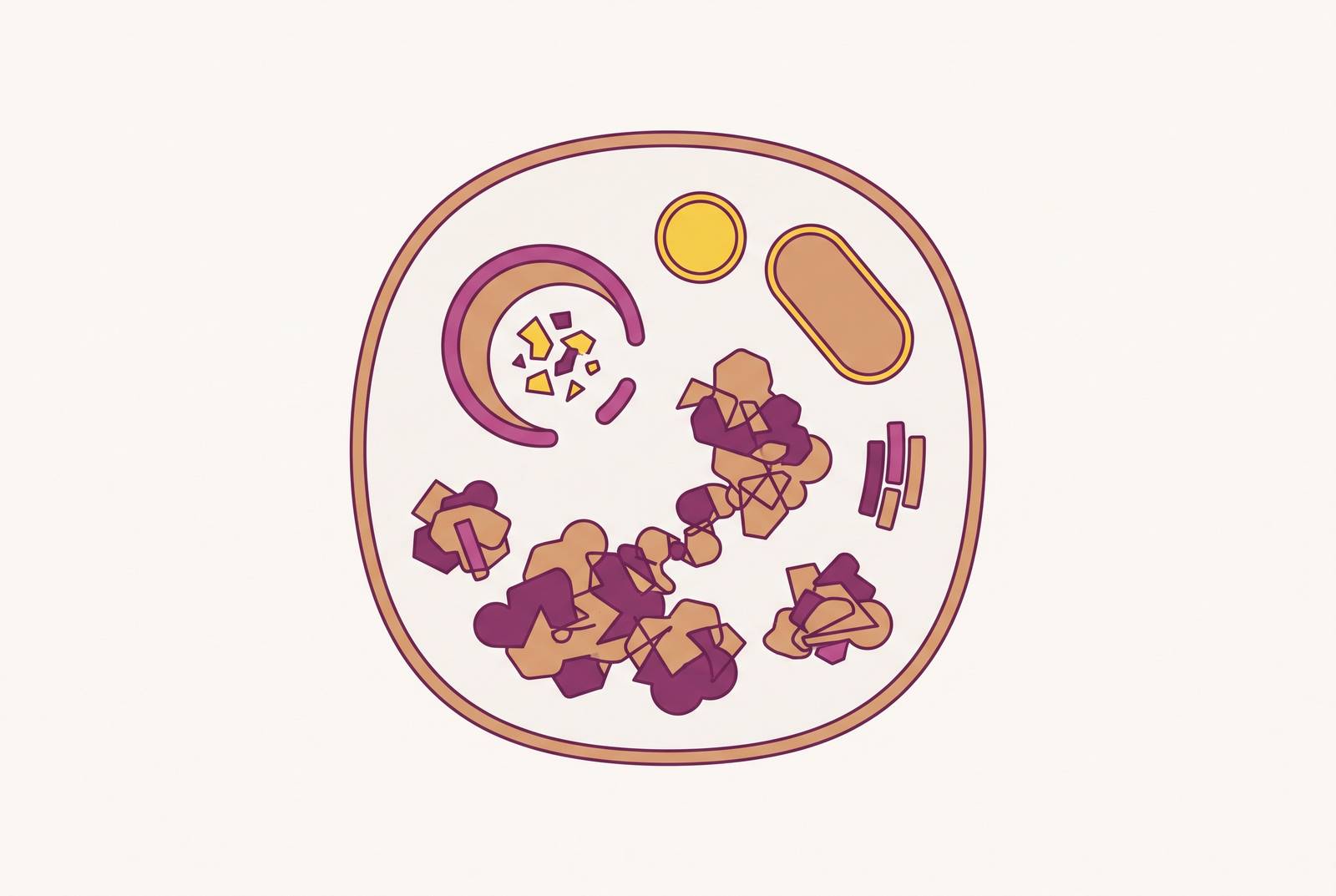
Cellular aging keeps circling back to one broken process: autophagy, the system cells use to clear out their own junk. When it slows down, the junk doesn't just sit there quietly. It builds into aggregates that jam the very machinery meant to remove them, and that feedback loop appears, in slightly different costumes, in heart failure, Alzheimer's, Parkinson's, and a good chunk of what gets filed under "normal aging."" Once you see the mechanism, it stops looking like an inevitability and starts looking like something with actual, measurable levers.
How autophagy works as the cell's quality-control system
Autophagy is a recycling process built into every cell, and its job is to break down and reuse worn-out parts: misfolded proteins, damaged mitochondria, stray lipid droplets, whatever's clogging things up. There are three known flavors, macroautophagy, microautophagy, and chaperone-mediated autophagy, but macroautophagy is the one with the deepest research base and the clearest tie to aging, so that's the focus here.
The process runs in stages. A structure called a phagophore forms first, then elongates into a double-membrane sac called an autophagosome, which wraps around the cargo it's collecting. That autophagosome then fuses with a lysosome, the cell's acid-filled breakdown chamber, forming an autolysosome where the actual degradation happens. Afterward, the lysosome gets reformed and the cycle starts again.
None of this runs on a handful of genes. More than 40 autophagy-related proteins coordinate the process, and their expression shifts depending on the tissue and the metabolic context. A neuron and a liver cell aren't running identical autophagy programs. That matters later, because it means "autophagy decline" is several dials, in several rooms, drifting out of sync. It's several dials, in several rooms, drifting out of sync.
What goes wrong with autophagy as the body ages
Autophagy declines with age. That much is settled. What's still being mapped out is exactly which molecular failures cause that decline and in what order, and that gap should be admitted honestly rather than glossed over. This is active science.
What makes the decline hard to reverse is that it doesn't fail at one step. It fails at several, simultaneously.
At initiation, ULK1 phosphorylation drops off, a protein called RUBCN gets upregulated and acts as a brake on autophagosome maturation, and VPS34 picks up acetylation marks that alter its activity. WIPI2 levels fall. Transcription of ATG2 and ATG9, both needed to get the process moving, declines too.
At elongation, cells lose ATG3, and increased disulfide bonding between ATG3 and ATG7 impairs the partnership those two proteins need to function. PKA-driven phosphorylation of LC3B at a site called Ser12 stops LC3B from attaching to the autophagosomal membrane the way it needs to.
Then there's the terminal stage, and this is arguably the costliest failure of the three. TFEB, the master regulator of lysosomal biogenesis, shows reduced activity with age, impairing the cell's ability to maintain an adequate lysosomal pool. The downstream capacity to actually degrade cargo diminishes as a result. Even when autophagosomes form just fine upstream, they arrive at a lysosomal system that can't finish the job. Cargo backs up.
Why does this hit some cells harder than others? Post-mitotic cells, neurons and cardiomyocytes especially, can't divide. A dividing cell at least has the option of distributing its accumulated burden across daughter cells. A neuron formed decades ago is stuck holding whatever it failed to clear, for the rest of its life.
How protein aggregates build up once autophagy slows
Once autophagic flux slows, misfolded and aggregated proteins pile up, and that's not some side effect. It's the direct, mechanical consequence of disabling the main cleanup crew.
Oxidative damage to proteins (chemical modifications caused by reactive oxygen species) plays a driving role here too, tying the aging redox environment directly into protein aggregation. Proteins get damaged, and the system meant to catch and clear damaged proteins is already running at reduced capacity. Two failures compound each other.
Normally, three separate quality-control systems back each other up, and they are the autophagy-lysosome pathway, the ubiquitin-proteasome system, and a family of molecular chaperones that help proteins refold correctly. All three decline with age. That should give anyone pause, because it means there's no redundant backstop left standing when one system falters. They're all falling at once.
A protein called p62, also known as SQSTM1, makes this feedback loop visible in a way few other markers do. Its job is to tag ubiquitinated protein aggregates and hand them off to autophagosomes for disposal. When autophagy is running well, p62 gets degraded right along with the cargo it delivers. When autophagy slows, p62 has nowhere to go, so it piles up alongside the very aggregates it failed to clear. That makes elevated p62 a reasonably direct proxy for impaired autophagic flux, and the numbers back it up: in aged skeletal muscle, p62-positive fibers show up in 14% of muscle fibers, compared to just 1% in young muscle. Fourteen times the burden, in the same tissue type, differing only by age.
The worst-hit organs: heart and brain
Both organs share a structural feature that makes them especially exposed: they're built mostly from post-mitotic cells. Cardiomyocytes and neurons can't divide their way out of a mess, so autophagy isn't just one tool among several for them, it's close to the only tool they've got for a lifetime of upkeep.
Research using primary adult cardiomyocytes from C57BL/6J mice confirmed something intuitive but important: contraction amplitude drops with age. Alongside that drop, autofluorescence increases, and that autofluorescence is a signature of lipofuscin, a fatty, undegradable pigment that builds up specifically because autophagy can't clear it.
The causal chain gets tested directly in that same line of research. Treating cardiomyocytes with artificial lipofuscin reduced both their contraction amplitude and their autophagic flux, and rapamycin (a drug that induces autophagy) mitigated the contractile dysfunction that lipofuscin caused. Then the experiment gets more precise: blocking autophagy at a late stage, using chloroquine or concanamycin A, reduced contraction amplitude, but blocking it at an early stage, using 3-methyladenine, didn't affect contraction within 24 hours, pointing to the lysosomal clearance step as the protective one. That's a fairly clean signal. The lysosomal clearance step, specifically, is what protects the heart, keeping cardiomyocytes contracting normally. It's the lysosomal clearance step, specifically, that keeps cardiomyocytes contracting normally. Lipofuscin doesn't just sit there looking inert. It actively impairs heart muscle function by choking off that late-stage flux.
The brain runs a parallel story with different named actors. Autophagy failure and aggregate buildup rank as a core hallmark of brain aging, and age itself remains the single biggest risk factor for both Alzheimer's disease and Parkinson's disease. Four aggregate types show up across the major neurodegenerative diseases: tau forms neurofibrillary tangles in Alzheimer's, alpha-synuclein forms Lewy bodies in Parkinson's, TDP-43 aggregates show up in ALS and frontotemporal dementia, and huntingtin aggregates define Huntington's disease. Different proteins, different diseases, but p62/SQSTM1 is present across multiple of these aggregate types. That's the shared upstream mechanism showing its fingerprints across four supposedly distinct diseases.
What happens after the aggregate forms is disease-specific: neuroinflammation, oxidative stress, mitochondrial dysfunction, synaptic failure, each cascade with its own particular flavor depending on which protein misfolded and where. But the origin point, a clearance system that can't keep up, looks the same across all four. mTOR signaling adds another layer specific to the brain: mTOR signaling has been linked to the regulation of both neurofibrillary tangle and amyloid-beta plaque pathology, connecting the metabolic regulation of autophagy to the pathology itself.
None of this means the heart and brain are the only organs that pay this price. They're the clearest illustration, because post-mitotic cells make the stakes visible fastest. Wherever cells can't divide their way to a clean slate, autophagy decline is going to leave a mark eventually.
Metabolic state, insulin, mTOR, and fasting control the autophagy dial
mTOR sits at the center of this whole system, functioning as a switch rather than a dimmer. High insulin and abundant amino acids keep mTORC1 turned on, and an active mTORC1 keeps autophagy suppressed, because the cell is in growth mode, not cleanup mode. When glucose and amino acids drop, mTOR deactivates, protein synthesis slows, and autophagy kicks in. It's a fairly elegant piece of cellular logic: build when resources are abundant, recycle when they're scarce.
Chronic obesity and metabolic syndrome throw a wrench into that logic. Persistent cellular stress in that state suppresses insulin-mediated autophagy over time, and that suppression eventually produces insulin resistance, which itself tends to climb with age and shows up alongside chronically elevated insulin levels. So the loop closes on itself: high insulin blocks autophagy, blocked autophagy contributes to resistance, resistance keeps insulin elevated. Heightened insulin accelerates aging partly through this exact route, pushing cells toward more biosynthesis and less clearance of the damaged parts that biosynthesis leaves behind.
Skeletal muscle offers one of the clearer windows into fixing this. Autophagy plays a direct role in how muscle cells handle glucose and respond to insulin, and exercise induces autophagy in muscle tissue while improving insulin sensitivity, a pattern with support across animal models. Moving the muscle improves both sides of the equation together.
And this metabolic thread loops right back to the brain. Impaired insulin and IGF-1 signaling are features of both ordinary brain aging and late-onset Alzheimer's disease. The mTOR story from this section and the neurodegeneration story from the last one aren't really two separate stories. They're the same failure, viewed from two different angles.
Interventions with evidence behind them and their effects on autophagy
Every intervention that holds up here works through one of the levers already laid out: suppressing mTOR, activating AMPK, switching on TFEB, or boosting lysosomal function directly. Once that's clear, it gets a lot easier to sort out which claims about "boosting autophagy" actually hold up.
Fasting and caloric restriction sit at the center of the evidence base. Dietary restriction, along with reduced insulin/IGF-1 signaling and reduced mTORC1 activity, extends both lifespan and healthspan in model organisms, and autophagy induction is at least part of why. Mechanically, nutrient scarcity deactivates mTOR, which frees up ULK1 to kick off autophagy, while AMPK gets activated in parallel, pushing the cell toward a broader stress-resistance program. This evidence base is strongest in worms, flies, and rodents. Human data specifically measuring autophagic flux under fasting protocols is still thin.
Exercise has a meaningful evidence base behind it on this particular point, including in humans. Combined with the earlier point about muscle insulin sensitivity, exercise ends up hitting both sides of the metabolism-autophagy relationship at once.
On the pharmacological side, five compounds occur repeatedly in longevity research, and all five converge, at least partly, on mTORC1 inhibition:
Rapamycin inhibits mTORC1 directly. It's the most studied compound in this group, inducing autophagy and lysosomal biogenesis through TFEB activation, and it's the same compound that mitigated lipofuscin-induced contractile dysfunction in the cardiomyocyte research described earlier. Metformin, a long-established metabolic drug, suppresses mTOR indirectly and carries autophagy-inducing effects as a byproduct of its glucose-lowering action. Spermidine, a naturally occurring polyamine, induces autophagy and has been studied in model organisms. Urolithin A, produced when gut bacteria metabolize certain polyphenols, promotes autophagy and has human data behind it in the context of muscle and mitochondrial health. Resveratrol activates autophagy indirectly, though the evidence for it sits mostly in model systems rather than humans.
Every one of those five shares mTORC1 inhibition as a mechanism, and that shared mechanism comes with a shared catch. mTOR doesn't just suppress autophagy, it also drives normal growth and protein synthesis. Suppress it broadly and indefinitely, and there's a real trade-off to weigh, not a free upgrade.
Biomarkers
The sources checked for this guide are listed below.