Intercellular Communication Breakdown in Aging Tissues
Aging spreads through organs when their communication network breaks down.
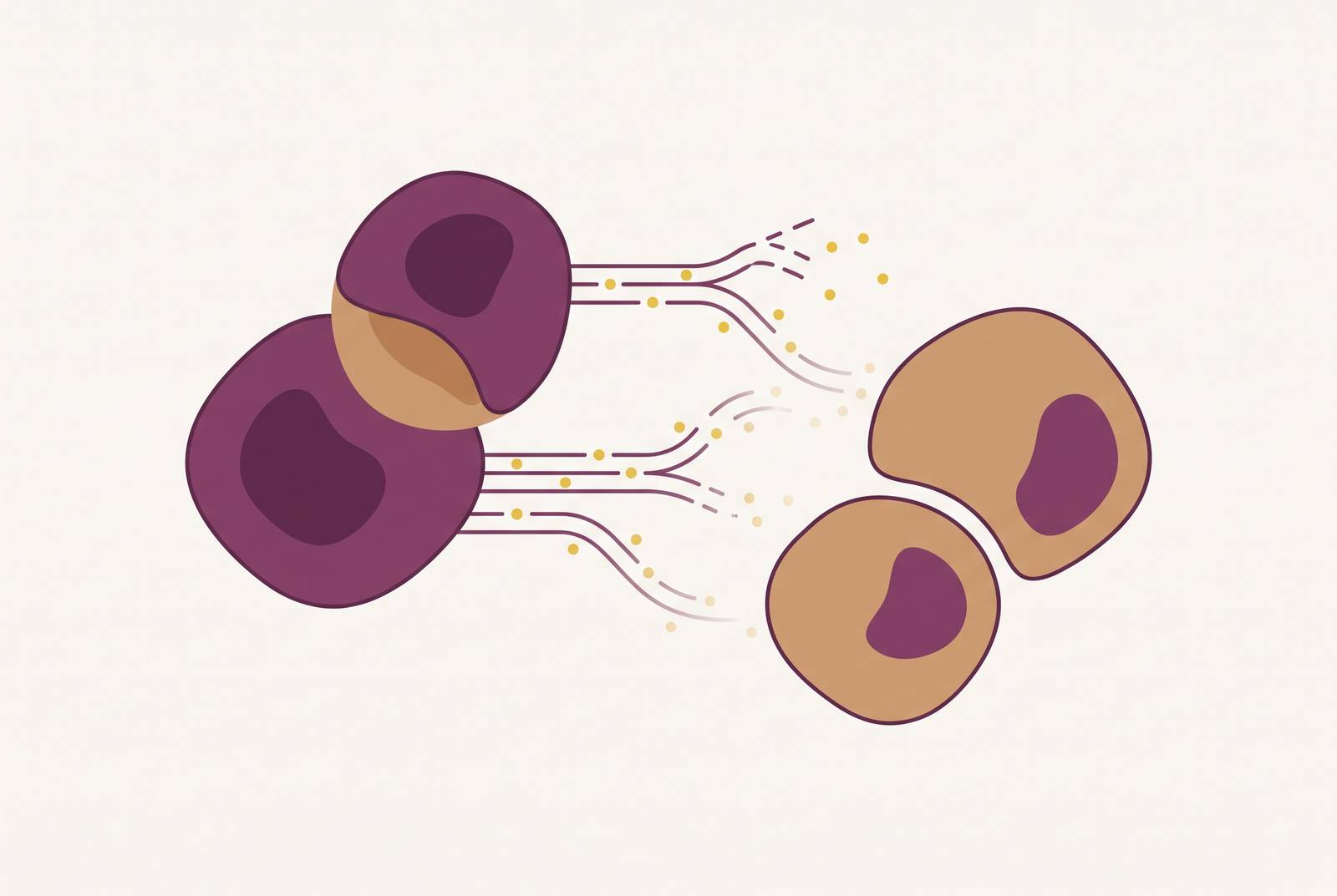
Aging doesn't happen because cells wear out one by one, like rust spreading across a car. It happens because the signaling network connecting those cells breaks down, so organs stop coordinating repair, metabolism, and immune defense the way they used to. That distinction explains something doctors have noticed for decades: heart disease, dementia, and metabolic decline tend to appear together, in the same person, in the same decade, rather than as isolated events.
A February 2026 review in Frontiers in Cell and Developmental Biology stated that organismal aging comes down to "the progressive breakdown of inter-organ homeostatic coordination rather than the decline of individual organ function." Not organs failing separately. A communication system failing collectively. Once you see aging this way, the clustering of age-related disease stops looking like bad luck and starts looking like the predictable output of a signaling network losing its ability to keep organs talking to each other.
Intercellular communication's place in the 12-hallmark framework of aging
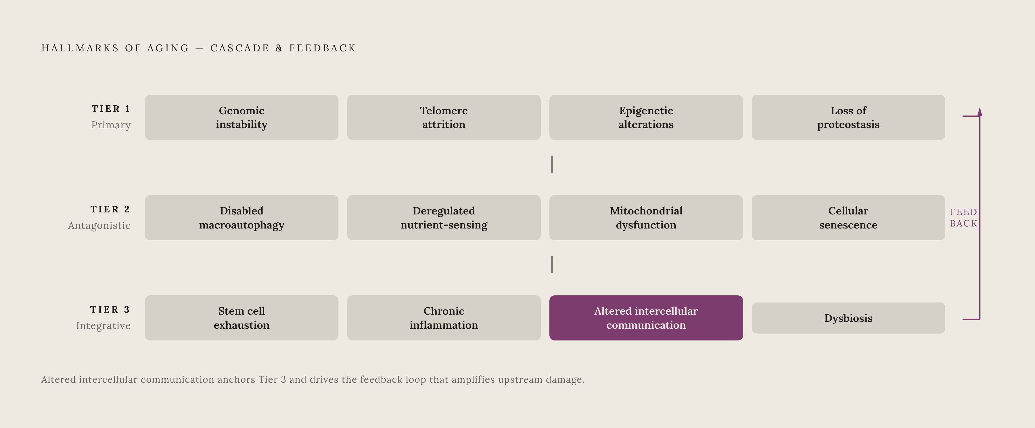
Researchers group the drivers of aging into 12 hallmarks, a framework cited in a 2025 Frontiers in Cell and Developmental Biology piece. The list: genomic instability, telomere attrition, epigenetic alterations, loss of proteostasis, disabled macroautophagy, deregulated nutrient-sensing, mitochondrial dysfunction, cellular senescence, stem cell exhaustion, altered intercellular communication, chronic inflammation, and dysbiosis.
These 12 sort into three layers. Primary hallmarks are raw damage accumulation: genomic instability, telomere attrition, epigenetic drift, and misfolded proteins piling up faster than cells can clear them. Antagonistic hallmarks are compensatory responses that start out helpful and eventually turn against the organism, things like deregulated nutrient sensing, mitochondrial dysfunction, and cellular senescence. Then there's the integrative layer: stem cell exhaustion, chronic inflammation, and altered intercellular communication. This is where damage shifts from local to systemic, because the signaling network that once coordinated repair, metabolism, and immune defense across cells has broken down.
That placement matters. Altered intercellular communication doesn't start the cascade, it sits downstream of it, a symptom of damage that's already accumulated elsewhere. But once the communication layer is compromised, it stops behaving like a passive symptom. It starts driving further deterioration across organ systems, feeding back into the very damage that caused it. That's a self-amplifying loop.
Why does this matter for anyone outside a research lab? Because it means intercellular communication breakdown is often the first thing that becomes visible, clinically, in blood work or symptoms, even though it's the last hallmark to activate. By the time someone's bloodwork shows the signature of communication failure, multiple upstream failures have already been compounding for years.
How senescent cells generate the SASP, turning local damage into a systemic signal
Cellular senescence is a stable cell cycle arrest. The cell stops dividing, but it doesn't die, and it doesn't stay quiet. It develops what's called the senescence-associated secretory phenotype, or SASP, a cocktail of pro-inflammatory factors the cell pumps into circulation on an ongoing basis: IL-6, IL-1α, TNF-α among them.
Under normal, younger physiology, the SASP serves a purpose. It's a flare signal, part of the normal short-term response to cellular damage. That's the intended design. But the SASP does something else at the same time: it propagates senescence-inducing signals to nearby cells, and even to cells in distant tissues, pushing otherwise healthy cells into secondary senescence. One damaged cell, left alone, doesn't stay one damaged cell.
Age changes the equation. Immune surveillance, the system responsible for hunting down and clearing senescent cells, declines with age. So senescent cells accumulate faster than the immune system can remove them. Instead of a short, resolved burst of inflammation (the kind that shows up during wound healing and then shuts off), the body ends up with a low simmer of SASP signaling that never resolves. Persistent, not acute. Chronic, not self-limiting.
Two pathways implicated in the inflammatory signaling inside senescent cells are CDC42 GTPase and NF-κB. Notably, knocking down CDC42 expression extended longevity in C. elegans, a finding that points to this pathway as a live target for intervention, not just a bystander in the process.
How mitochondria function as inter-organ communication hubs
Mitochondria used to get described as the power plants of the cell, tidy little energy factories doing their job inside a single cell's walls. That picture is incomplete. Mitochondria form dynamic, interconnected networks, and their architecture, quality control, and communication with mitochondria in other cells shape whole-body homeostasis.
How do mitochondria talk to each other across cells? A few mechanisms stand out. Tunneling nanotubes act as direct physical bridges between cells. Extracellular vesicles ferry mitochondrial cargo from one location to another. Cells can undergo transient fusion. And mitochondria release circulating mitokines and metabolites that travel through the bloodstream and act on distant tissue.
The liver functions as something like a central relay station in this network. Hepatic mitochondria absorb metabolic and inflammatory stress signals and broadcast them systemically through three main channels: mitokines driven by the mitochondrial unfolded protein response (FGF21 and GDF15 are the named examples), metabolites generated by mitochondrial metabolic reprogramming, and danger signals like mitochondrial reactive oxygen species and oxidized mitochondrial DNA. These signals travel outward to skeletal muscle, adipose tissue, the brain, and the heart, reshaping how those organs handle energy metabolism and mitochondrial function.
So what happens when this network degrades with age? The liver keeps sending signals, but the signals shift character, from routine regulatory chatter toward stress and danger signaling. Downstream organs receive a steady diet of alarm bells instead of coordination cues. That's a different kind of failure than a single organ simply running out of energy.
Exosomes and extracellular vesicles as the corrupted messaging layer
Extracellular vesicles, EVs for short, and their smaller subset, exosomes, are nano-sized packages cells release to move cargo, proteins, lipids, and mRNAs, across tissues and even across biological barriers that would otherwise block direct contact. Think of them as sealed envelopes carrying instructions from one part of the body to another.
Under normal conditions, EVs help coordinate tissue repair, keep immune activity in balance, and support metabolic regulation across organ systems. They're part of the infrastructure, not an afterthought.
Age changes both how many EVs circulate and what they're carrying. Research has linked age-related shifts in EV abundance and cargo composition to impaired communication across organ systems. A finding from nematode research makes the point sharply: the cargo and context of vesicle release shift dramatically with age, such that the same biological vehicle can have opposite effects depending on the cellular environment sending it out.
That finding should reframe how anyone thinks about EVs. They're not neutral carriers doing the same job regardless of context. As senescent cells and dysfunctional mitochondria come to dominate more of the tissue environment, the cargo loaded into these vesicles shifts too, and what gets delivered downstream starts working against the recipient tissue instead of for it.
The inflammatory markers that make this cascade visible in a blood test
A June 2026 review in PMC found that traditional inflammatory markers "fail to capture the complexity, tissue specificity, and causal architecture of inflammaging." Traditional inflammatory markers fail to capture the complexity, tissue specificity, and causal architecture of inflammaging. These markers are still what's available on a standard blood panel today, and understanding what they actually measure matters.
High-sensitivity C-reactive protein, hs-CRP, gets made by the liver in response to inflammation. It picks up low-level systemic inflammation, the kind that causes no symptoms but may be quietly working on blood vessels and organs in the background. It's a downstream readout of SASP activity and inflammaging broadly.
IL-6 sits closer to the source. It's one of the primary components of the SASP itself, and one of the most studied cytokines in inflammaging research. It's not on a standard panel, but specialized testing increasingly makes it available, and it offers a more direct window into senescent cell burden than CRP alone. In the PolSenior study, which followed 3,496 participants aged 65 and older, IL-6 rose in an age-dependent pattern (p<0.001) and ran lower in people classified as aging successfully compared to the rest of the cohort (p<0.001). IL-6 tracked with age-dependent deterioration across that population.
CRP showed a similar pattern in the same cohort, among 3,632 participants: age-dependent increase (p=0.003), lower in successful agers (p<0.001), predictive of both performance and mortality.
The neutrophil-to-lymphocyte ratio, NLR, comes free with a standard complete blood count, no extra test needed, and is used as a marker of immune imbalance and systemic inflammation.
Homocysteine deserves particular attention, because it's one of the most actionable markers on this list. Elevated homocysteine predicts cardiovascular disease, cognitive decline, and all-cause mortality, and levels respond directly to B12, B6, and folate status, nutrients that are simple to correct. Optimal sits below 8 µmol/L. Yet many labs don't flag anything up to 15 µmol/L as abnormal, despite evidence that harm accumulates above 10 µmol/L. The next time a result comes back "normal," that gap between the lab's reference range and what the science calls optimal remains."
Putting these together, a pattern emerges. SASP-driven secretion of IL-6, TNF-α, and IL-1α pushes hs-CRP and related markers upward systemically. So someone showing elevated hs-CRP alongside elevated IL-6 is likely carrying a heavier senescent cell burden. None of these tests are exotic. Most sit on standard or low-cost panels already. The value isn't in ordering something rare, it's in knowing what the numbers mean once inflammaging, not infection, becomes the more likely explanation.
Senescence-specific biomarkers at the frontier of what can be measured today
IL-6, TNF-α, and CRP measure inflammation in general. They can't distinguish SASP-driven inflammaging from an active infection or an autoimmune flare. That's the ceiling on what standard bloodwork can currently reveal, and it's why researchers keep pushing for tools that measure senescent cells more directly.
A few approaches are further along than others. The SA-β-Gal assay measures senescence-associated beta-galactosidase activity, a well-established lab signature of senescent cells. Cell cycle checkpoint proteins, p16 and p21, offer another angle, since senescent cells rely on these checkpoints to stay permanently arrested. Cell morphology assessment rounds out the toolkit. None of these are blood tests you'd get at a routine physical, but together they give a more direct read on senescent cell burden itself.
p16INK4a, encoded by the CDKN2A locus, stands out as a particularly promising signal, and it produces several variant transcripts in humans. In a clinical trial testing the senolytic combination dasatinib plus quercetin (D+Q) in postmenopausal women, researchers looked at effects on bone metabolism. Women in the highest tertile for T-cell expression of one particular variant, p16_variant 5, showed the strongest skeletal response to treatment. That's a meaningful clue: p16 variants might eventually work as a selection tool, helping identify which patients stand to benefit most from senolytic therapy before it's given.
None of this belongs in routine clinical practice yet. These remain research tools, used in trials rather than in a standard workup. A July 2026 article in PMC named the gap directly, pointing to an "increasing need for biomarkers of senescent cell burden to facilitate the selection of participants for clinical trials." The tools that would let clinicians measure senescence directly, rather than inferring it from inflammatory proxies, are still being built. What's clear already is the direction: senescent cell burden, once it can be measured cleanly, becomes the variable that decides who actually responds to the next generation of anti-aging treatment.
Sources
- Frontiers | Hepatic mitochondrial signaling as a systemic hub: inter-organ communication networks in aging and aging-related diseases
- Communication breakdown: senescent cells in interorgan communication of aging: Trends in Endocrinology & Metabolism
- Mitochondrial Network Dynamics in Aging: Cellular Mechanisms, Intercellular Communication, and Their Impact on Tissue Adaptability | MDPI
- pmc.ncbi.nlm.nih.gov
- The Hallmarks of Aging: From Molecular Mechanisms to Clinical Translation
- pmc.ncbi.nlm.nih.gov
- fightaging.org
- ncbi.nlm.nih.gov